




















_800.png "No Hoof, No Horse: Equine Hoof Anatomy | Equine Science Matters™")
We have probably all heard the phrase ‘No Hoof, No Horse’ but do you know where it started? Or how did it come to be?
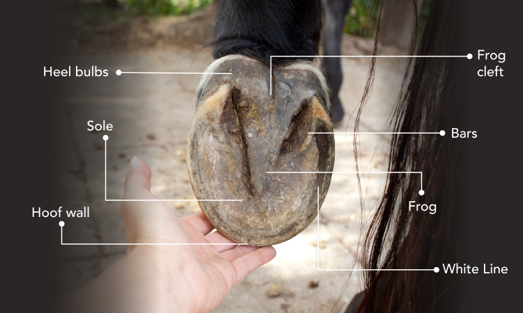
The horse’s hoof is a complex structure that plays an important role in absorbing shock, providing balance and supporting the horse’s weight. The external structures of your horse’s hoof that we see every day also play key roles in many internal systems. These structures are:
-
Hoof wall
-
Frog + Frog cleft
-
Bars
-
White line
-
Hoof wall
-
Sole
-
Coronary band
-
Heel bulbs
Hoof wall
The hoof wall is made up of Keratin, a tough fibrous protein, and is key to supporting your horse’s weight, maintaining balance and stability.
The Frog
The frog is an important part of the shock absorbing complex of the hoof, and also works as part of your horse’s circulatory system, aiding blood flow in your horses’ limbs, but how does the frog do this?
When the hoof is weight-bearing (on the ground), the energy created by the force of the hoof meeting the ground is dissipated sideways, leading the frog and underlying digital cushion to expand sideways, both dissipating the pressure and filling these structures with blood. Subsequently, when the hoof is lifted from the ground, the frog contracts, delivering blood back up the leg towards the heart whilst simultaneously dissipating shock. Think of the frog as a water pump; when you draw the handle up, creating pressure, water is drawn towards the pump and spout. When the handle is pushed down, releasing pressure, the water returns to its source.
The Bars, Sole and White line
The bars are the protruding lines, found on either side of your horse’s frog, that provide balance and aid weight bearing.
The sole plays a large role in the protection of the hoofs internal structure, this is aspect of the hoof that we often pay the most attention to with flaking or crumbling soles being a common sign spotted by horse owners that their horses’ hooves may need support. Comprised of a keratinized tissue, this aspect provides the strength to absorb the shock placed on the limbs in addition to protection and support against concussive forces.
The white line is the junction where the hard outer hoof wall meets the softer, inner sole. The point where the sensitive and insensitive laminae meet and the place that ‘white line disease’ or ‘Seedy toe’ begins to attack your horse’s hoof. Making sure that this area of the hoof is structurally sound and stable helps prevent the invasions of bacteria or fungi that cause these diseases and affect the horses entire hoof stability long term.
The coronary band and heel bulbs
Whilst these structures are often overlooked when you are checking your horses’ hooves during your daily hoof picking, they are integral to new growth and impact absorption.
Acting as the ‘lifeblood’ of the hoof, the coronet (or coronary) band is the critical junction where the skin of the lower leg meets the hard hoof capsule. The coronet is responsible for about 70% of new hoof wall growth, producing keratinized cells for 6 to 8mm of growth per month. This area is also packed with digital nerve branches and blood vessels, making it highly sensitive to pressure, aiding circulation.
While the heel bulbs are rounded and allow the hoof to contract and expand, as your horse moves, absorbing impact and maintaining balance. When shoeing a horse, your farrier will consider this action of the heel bulbs to prevent restricting this important mechanism.
The hooves inner structures
-
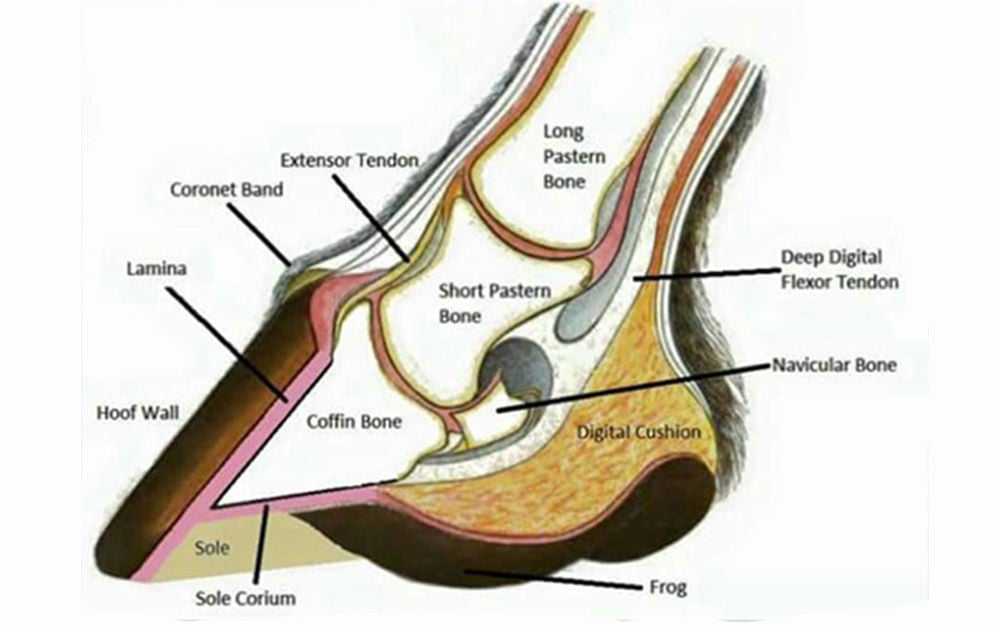
The pedal bone (third phalanx or P3) is the main bone within the hoof and provides attachment for important tendons and ligaments.
-
The collateral cartilages, located on either side of the bony structures within the hoof, help with expansion and contraction, allowing flexibility and helping maintain a smooth range of motion.
-
The navicular bone works alongside the navicular bursa, a fluid-filled sac that reduces friction between the navicular bone and the deep digital flexor tendon (DDFT) during movement.
-
Positioned beneath the frog is the digital cushion, another important shock absorber that helps reduce concussion travelling up through the joints of the limb.
-
The pedal bone is suspended within the hoof by the laminae—thousands of interlocking, collagen-rich structures that securely attach the hoof wall to the bone while providing both strength and flexibility.
This is the site where laminitis occurs, and is the inflammation of the laminae, weakening the bond between the hoof wall and the pedal bone. Maintaining the health of the laminae is extremely important, as it bonds the pedal bone inside the hoof to the structure of the hoof wall. If the laminar tissue loses its ability to function correctly, there can be large and painful changes to the internal structure of the hoof. In severe cases, the pedal bone may rotate or sink within the hoof capsule, making this one of the most painful conditions affecting horses.
Signs to watch for include:
-
Lameness.
-
Leaning back onto the heels to relieve pressure on the painful toe.
-
Frequently shifting weight between feet.
-
Increased digital pulses.
Common causes include inflammatory diseases such as certain types of colic, endocrine disorders including Cushing's disease (PPID) and EMS, steroid use, mechanical overload, and diet-related factors.
Additional Problems associated with the hoof:
Osteoarthritis
The interphalangeal, the most weight bearing bone, is under constant concussion. Factors like age and trauma will increase the likelihood of osteoarthritis, causing cartilage wear, synovitis, and bony changes.
Navicular Disease
Navicular disease is a chronic, degenerative condition affecting the navicular apparatus. Repeated concussion can lead to degeneration of the fibrocartilage, inflammation of the navicular bursa, and changes to the surrounding hoof structures, resulting in persistent heel pain.
White line disease
Also known as seedy toe, white line disease occurs when the hoof wall begins to separate from the laminae at the white line, creating cavities that allow bacteria and fungi to invade the hoof.
Thrush
Thrush is a common bacterial infection that primarily affects the frog, more specifically the soft frog. The warm and damp conditions that are easily created in the cleft of the frog are ideal for bacteria to proliferate, and can cause foul-smelling discharge and deterioration of the frog tissue that are usually referred to as thrush.
Contracted heels
Contracted heels occur when the back of the hoof narrows abnormally, restricting the natural expansion of the hoof. This can lead to compression of the frog and heel bulbs and is commonly associated with poor hoof balance or incorrect shoeing.
In Conclusion
The saying "No Hoof, No Horse" perfectly summarises just how important healthy hooves are to a horse's overall wellbeing.
Damage, imbalance or disease within the hoof can alter the horse's way of going, reduce its ability to absorb shock, and place excessive strain on the joints and soft tissues of the limb. By becoming familiar with both the condition and shape of your horse's hooves, you can identify subtle changes early, helping to prevent more serious problems that may lead to poor performance or lameness.