


 Ph.D. Equine Nutrition")

Anouk Frieling, MSc Equine Sciences, BSc (Hons)
Atypical myopathy (AM) is a seasonal, non-exercise related, disease causing acute rhabdomyolysis (rapid breakdown of damaged muscle tissue) which affects the muscles of the horse
Atypical Myopathy Causes and Pathophysiology
Equine atypical myopathy mainly affects the respiratory muscles and muscles that support the skeleton and maintain balance
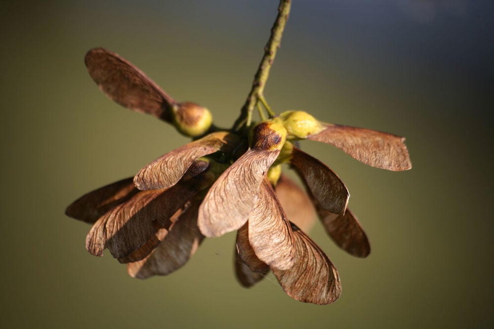
Figure 1: During autumn the seeds from the sycamore tree fall on the pasture on which horses graze. These seeds can be toxic and horses ingesting these seeds can develop atypical myopathy.
After ingestion, HGA interferes with metabolic processes
Clinical signs and Diagnosis
Clinical signs often start with the sudden onset of muscle weakness and stiffness
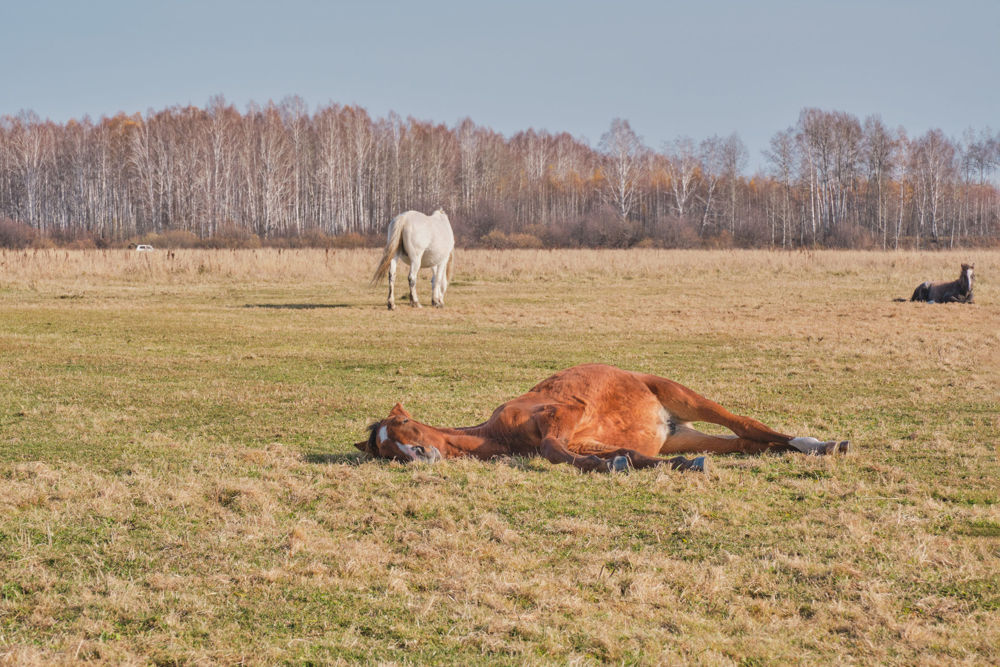
Figure 2: Atypical Myopathy affects the muscles of the horse. Horses that lie down and do not stand up is one of the clinical signs of AM.
Diagnosing AM based on the clinical signs is difficult and can often lead to a wrong diagnosis as the clinical signs are very similar to those of other equine diseases
Treatment
As mentioned, currently there is no specific treatment for AM affected horses
One of the symptoms of AM is secreting dark urine which is a signs of dehydration. Therefore, providing fluids is essential for rehydration, and will enhance renal clearance of excess metabolites and correction of blood parameters
As previously discussed, due to AM the fatty acid metabolism in muscle tissue is disturbed and will shift from aerobic to anaerobic
Studies suggest that the supplementation of vitamin E, selenium, carnitine and riboflavin can support AM affected horses and can increase chances of survival
Prevention
Preventing AM is important as currently there is no specific treatment. Firstly, to prevent AM in grazing horses, ingestion of toxic sycamore seeds or spouts should be prevented by checking the environment of the horse. If found, seeds and sprouts should be removed from the horse’s environment
Conclusion
Atypical myopathy is an acute seasonal disease in grazing horses, affecting the muscles of the horse. The onset of the disease is caused due to ingestion of sycamore seeds, sprouts or leaves which contain the toxic compound HGA. After intoxication the disease progresses rapidly and is therefore highly fatal. Clinical signs include muscle weakness and stiffness but the signs of the disease are very similar to other muscle diseases in horses, making it difficult to correctly diagnose a horse with AM. Currently, there is no specific treatment for the disease as the full pathophysiology is still unknown. Available treatment is mainly supportive to be able to increase the chances of survival for affected horses. Because there is no treatment available, it is important to implement good pasture management and prevent horses from ingesting toxic composites.
References
Bochnia, M., Ziegler, J., Sander, J., Uhlig, A., Schaefer, S., Vollstedt, S., Glatter, M., Abel, S., Recknagel, S., Schusser, G. F., Wensch-Dorendorf, M., & Zeyner, A. (2015). Hypoglycin a content in blood and urine discriminates horses with atypical Myopathy from clinically normal horses grazing on the same pasture. PLoS ONE, 10(9): 1-15.
Brandt, K., Hinrichs, U., Glitz, F., Landes, E., Schulze, C., Deegen, E., Pohlenz, J., & Coenen, M. (1997). Atypische myoglobinurie der weidepferde. Pferdeheilkunde, 13(1): 27-34.
Cassart, D., Baise, E., Cherel, Y., Delguste, C., Antoine, N., Votion, D., Amory, H., Rollin, F., Linden, A.,Coignoul, F., & Desmecht, D. (2007). Morphological alterations in oxidative muscles and mitochondrial structure associated with equine atypical myopathy. Equine Veterinary Journal, 39(1): 26-32.
Fabius, L. S., & Westermann, C. M. (2018). Evidence-based therapy for atypical myopathy in horses. Equine Veterinary Education, 30(11): 616-622.
Fagan, M. M., Pazdro, R., Call, J. A., Abrams, A., Harris, P., Krotky, A. D., & Duberstein, K. J. (2017). Assessment of oxidative stress and muscle damage in exercising horses in response to level and form of vitamin E. Journal of Equine Veterinary Science, 52: 80-81.
Finno, C. J., Valberg, S. J., Wünschmann, A., & Murphy, M. J. (2006). Seasonal pasture myopathy in horses in the midwestern United States: 14 Cases (1998-2005). Journal of the American Veterinary Medical Association, 229(7): 1134-1141.
González-Medina, S. (2015). Update on the cause of equine atypical myopathy. The Veterinary Record, 176(6): 143-145.
González-Medina, S., Hyde, C., Lovera, I., & Piercy, R. J. (2021). Detection of hypoglycin A and MCPA-carnitine in equine serum and muscle tissue: Optimisation and validation of a LC-MS–based method without derivatisation. Equine Veterinary Journal, 53(3): 558-568.
González-Medina, S., Ireland, J. L., Piercy, R. J., Newton, J. R., & Votion, D. M. (2017). Equine atypical myopathy in the UK: Epidemiological characteristics of cases reported from 2011 to 2015 and factors associated with survival. Equine Veterinary Journal, 49(6): 746-752.
Harris, P., & Whitwell, K. (1990). Atypical myoglobinuria alert. Veterinary Record, 127(24): 603.
Herrera, C. M., Jordano, P., Guitián, J., & Traveset, A. (1998). Annual variability in seed production by woody plants and the masting concept: Reassessment of principles and relationship to pollination and seed dispersal. American Naturalist, 152(4): 576-594.
Karlíková, R., Široká, J., Jahn, P., Friedecký, D., Gardlo, A., Janečková, H., Hrdinová, F., Drábková, Z., & Adam, T. (2016). Equine atypical myopathy: A metabolic study. Veterinary Journal, 216: 125-132.
Karlíková, R., Široká, J., Mech, M., Friedecký, D., Janečková, H., Mádrová, L., Hrdinová, F., Drábková, Z., Dobešová, O., Adam, T., & Jahn, P. (2018). Newborn foal with atypical myopathy. Journal of Veterinary Internal Medicine, 32(5): 1768-1772.
Kranenburg, L. C., Westermann, C. M., de Sain-van der Velden, M. G. M., de Graaf-Roelfsema, E., Buyse, J., Janssens, G. P. J., van den Broek, J., & van der Kolk, J. H. (2014). The effect of long-term oral L-carnitine administration on insulin sensitivity, glucose disposal, plasma concentrations of leptin and acylcarnitines, and urinary acylcarnitine excretion in warmblood horses. Veterinary Quarterly, 34(2): 85-91.
Lemieux, H., Boemer, F., van Galen, G., Serteyn, D., Amory, H., Baise, E., Cassart, D., van Loon, G., Marcillaud-Pitel, C., & Votion, D. M. (2016). Mitochondrial function is altered in horse atypical myopathy. Mitochondrion, 30: 35-41.
Mizock, B. A., & Falk, J. L. (1992). Lactic acidosis in critical illness. Critical Care Medicine, 20(1): 80-93.
Palencia, P., & Rivero, J. L. L. (2007). Atypical myopathy in two grazing horses in northern Spain. Veterinary Record, 161(10):
Unger, L., Nicholson, A., Jewitt, E. M., Gerber, V., Hegeman, A., Sweetman, L., & Valberg, S. (2014). Hypoglycin A Concentrations in Seeds of Acer Pseudoplatanus Trees Growing on Atypical Myopathy-Affected and Control Pastures. Journal of Veterinary Internal Medicine, 28(4): 1289-1293.
van Galen, G., Marcillaud Pitel, C., Saegerman, C., Patarin, F., Amory, H., Baily, J. D., Cassart, D., Gerber, V., Hahn, C., Harris, P., Keen, J. A., Kirschvink, N., Lefere, L., Mcgorum, B., Muller, J. M. V., Picavet, M. T. J. E., Piercy, R. J., Roscher, K., Serteyn, D., … Votion, D. M. (2012). European outbreaks of atypical myopathy in grazing equids (2006-2009): Spatiotemporal distribution, history and clinical features. Equine Veterinary Journal, 44(5): 614-620.
Verheyen, T., Decloedt, A., de Clercq, D., & van Loon, G. (2012). Cardiac Changes in Horses with Atypical Myopathy. Journal of Veterinary Internal Medicine, 26(4): 1019-1026.
Votion, D. M., François, A. C., Kruse, C., Renaud, B., Farinelle, A., Bouquieaux, M. C., Marcillaud‐pitel, C., & Gustin, P. (2020). Answers to the frequently asked questions regarding horse feeding and management practices to reduce the risk of atypical myopathy. Animals, 10(2): 365.
Votion, D. M., Linden, A., Delguste, C., Amory, H., Thiry, E., Engels, P., van Galen, G., Navet, R., Sluse, F., Serteyn, D., & Saegerman, C. (2009). Atypical myopathy in grazing horses: A first exploratory data analysis. Veterinary Journal, 180(1): 77-87.
Votion, D. M., Linden, A., Saegerman, C., Engels, P., Erpicum, M., Thiry, E., Delguste, C., Rouxhet, S., Demoulin, V., Navet, R., Sluse, F., Serteyn, D., Van Galen, G., & Amory, H. (2007). History and clinical features of atypical myopathy in horses in Belgium (2000-2005). Journal of Veterinary Internal Medicine, 21(6): 1380-1391.
Votion, D. M., & Serteyn, D. (2008). Equine atypical myopathy: A review. Veterinary Journal, 178(2): 185–190.
Votion, D. M. (2012). The Story of Equine Atypical Myopathy: A Review from the Beginning to a Possible End. ISRN Veterinary Science, 2012: 1–14
Westermann, C. M., Dorland, L., Votion, D. M., de Sain-van der Velden, M. G. M., Wijnberg, I. D., Wanders, R. J. A., Spliet, W. G. M., Testerink, N., Berger, R., Ruiter, J. P. N., & van der Kolk, J. H. (2008). Acquired multiple Acyl-CoA dehydrogenase deficiency in 10 horses with atypical myopathy. Neuromuscular Disorders, 18(5): 355-364.
Westermann, C. M., van Leeuwen, R., van Raamsdonk, L. W. D., & Mol, H. G. J. (2016). Hypoglycin A Concentrations in Maple Tree Species in the Netherlands and the Occurrence of Atypical Myopathy in Horses. Journal of Veterinary Internal Medicine, 30(3): 880-884.
Wüger, C., Straub, R., & Gerber, V. (2006). Intravenöse flüssigkeitstherapie beim pferd. Pferdeheilkunde, 22(3): 327-336.
Zimmerman, J. L., & Shen, M. C. (2013). Rhabdomyolysis. Chest, 144(3): 1058- 1065.
Żuraw, A., Dietert, K., Kühnel, S., Sander, J., & Klopfleisch, R. (2016). Equine atypical myopathy caused by hypoglycin A intoxication associated with ingestion of sycamore maple tree seeds. Equine Veterinary Journal, 48(4): 418-421.