


 Ph.D. Equine Nutrition")

Nicola Menzies-Gow, MA VetMB, PhD, DipECEIM, CertEM(Int.med), FHEA, FRCVS, Professor of Equine Medicine, Royal Veterinary College.
Laminitis is now considered to be a syndrome associated with systemic disease (sepsis or endocrine disease) or altered weight bearing rather than being a disease itself (Patterson-Kane et al., 2018). There are three forms of laminitis, each of which occurs in association with very different diseases and occurs following very different sequences of events in the body:
SEPSIS-ASSOCIATED LAMINITIS
Sepsis-associated laminitis occurs secondary to a severe systemic (involving the whole body) inflammatory response and/or sepsis (bacterial infection in the blood) and so occurs in animals with, for example, severe gastrointestinal disease, pleuropneumonia (bacterial infection of the lungs that has extended into the pleural space) and septic metritis (bacterial infection of the uterus) following retention of the placenta. Toxins produced by the bacteria are absorbed into the blood stream where they set off a severe inflammatory reaction. Whilst there is evidence of inflammation in several places throughout the body such as the skin (Riggs et al., 2009), lungs and the liver (Stewart et al., 2009), the foot appears to be most severely affected with laminitis being the end result.
ENDOCRINOPATHIC LAMINITIS
Endocrinopathic laminitis is the commonest form of laminitis, accounting for 90% of cases of laminitis in some studies (Karikoski et al., 2011). It includes laminitis associated with abnormal regulation of the hormone insulin (insulin dysregulation; ID), as occurs in equine metabolic syndrome (EMS) and in a subset of animals with pituitary pars intermedia dysfunction (PPID; equine Cushing’s disease) (Durham et al., 2019). Exactly how insulin causes laminitis has yet to be fully determined. The current theory with the most supporting evidence is that insulin binds to and activates the insulin like growth factor-1 (IGF-1) receptor in the lamellae resulting in laminitis.
SUPPORTING LIMB LAMINITIS
Supporting limb laminitis (SLL) is uncommon (Wylie et al., 2015). However, it is a major contributor to treatment failure in painful limb conditions such as fractures and joint/tendon sheath infections as laminitis develops in the contralateral limb due to excessive weight bearing. It is thought that the increased weight bearing results in decreased blood supply to the foot as the blood supply is reliant on movement to help pump it around the foot. The decreased blood supply means that the delivery of oxygen and nutrients to the foot is reduced resulting in laminitis.
SYMPTOMS OF LAMINITIS
The symptoms of laminitis are the same regardless of type and include any combination of:
• Lameness that usually affects two or more limbs, but lameness in one limb is possible
• Reluctance to walk with a short, stilted gait and difficulty turning that is worse on hard ground
• Increased (or bounding) digital pulses
• Increased hoof wall temperature
• Weight shifting
• Pain on hoof tester pressure at the toe region just in front of the point of the frog
• Characteristic stance of leaning back on the heels and taking weight off the painful toe region
• Palpable depression at the coronary band
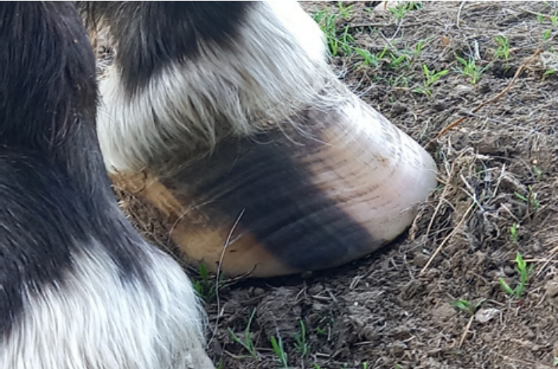
Figure 1. Divergent hoof growth rings are a sign of changes within the hoof associated with laminitis. Credit: Professor Menzies-Gow
The lameness can vary in severity from that which is only perceptible at the trot, through to spending prolonged periods recumbent. Some episodes of laminitis are subclinical and whilst there is no obvious lameness (or any other clinical sign), it subsequently becomes apparent as divergent hoof growth rings (Patterson-Kane et al., 2018) (Figure 1).
DIAGNOSIS OF LAMINITIS
A diagnosis of laminitis is usually based on the history, as it is often recurrent, and the clinical signs. X-rays are taken in cases where rotation and/or sinking of the pedal bone within the hoof capsule is suspected. Further diagnostic tests are performed to identify the presence of an underlying endocrine (hormone) disease in suspect animals. A diagnosis of EMS is made based on demonstrating the presence of ID. Insulin dysregulation can manifest in three ways and tests are required to identify each manifestation. High circulating insulin concentrations are detected by measuring blood insulin concentrations. Excessive release of insulin into the blood following ingestion of carbohydrate is identified using an oral sugar (OST) or glucose (OGT) test. An abnormal response of the tissues to insulin is detected using an intravenous insulin tolerance test. A diagnosis of PPID is made by measuring blood concentrations of the pituitary-derived hormone adrenocorticotrophic hormone (ACTH) and/or using a thyrotropin releasing hormone (TRH) stimulation test.
TREATMENT
Treatment involves providing pain relief and supporting the foot to prevent any/further movement of the pedal bone within the hoof. The underlying endocrine disease or primary condition causing sepsis-associated or supporting-limb laminitis needs to be treated specifically. Additionally, cryotherapy (cold therapy) is indicated in certain circumstances.
PAIN RELIEF
Non-steroidal anti-inflammatory drugs (NSAIDs) such a flunixin and phenylbutazone (bute) are the first choice for pain relief and there is no evidence to suggest that any one specific NSAID is superior to the next (Menzies-Gow et al., 2010). Although not a strong painkiller on its own, paracetamol appears to have an excellent effect in laminitic cases when used in addition to other NSAIDs (West et al., 2011). If NSAIDs do not provide enough pain relief, then opiates can be used in addition e.g., butorphanol, pethidine and morphine. Fentanyl patches applied directly to the skin can also be considered as it was found to be effective in horses with pain refractory to NSAID analgesia in one small clinical report (Thomasy et al., 2004). However, uptake of fentanyl from a skin patch is highly variable in adult horses (Orsini et al., 2006). Tramadol has also been advocated as it decreased forelimb offloading in four cases of chronic laminitis (Guedes et al., 2016), but current evidence does not support its use alone as it has a low absorption (~9%) from the gastrointestinal tract and it only lasts a very short time (~2 hours) (Stewart et al., 2011). If single drugs do not provide adequate analgesia, then several drugs can be used in combination in the hospital setting. Possible combinations typically involve NSAID administration in combination with an intravenous infusion up to four other types of drugs. A nerve-related component to the pain associated with laminitis has been demonstrated, making the drugs ketamine and gabapentin potentially suitable drugs. In one study, the combination of tramadol and ketamine resulted in decreased forelimb off-loading frequency and increased forelimb loading in horses with naturally occurring laminitis (Guedes et al., 2012). Gabapentin improved hindlimb pain that was probably associated with femoral nerve damage in one horse (Davis et al., 2007) and had no apparent adverse effects following oral administration in horses (Terry et al., 2010). The drug(s) that will provide sufficient pain relief in any individual horse is very variable and often it is a matter of trialling each drug to see the clinical response.
FOOT SUPPORT
Supporting the foot is essential. Additional support should be applied to the back two thirds of the foot to provide pain relief and to minimise the mechanical forces on the laminae and hence prevent/reduce pedal bone movement. The simplest method is to increase the depth of the bedding, ensuring that the bedding extends to the door where the horse will spend most of its day. Alternatively, or additionally, extra support can be applied directly to the foot itself using methods that can be broadly divided into frog only supports and combined frog and sole supports. There is no evidence to suggest that any one foot support method is superior (Menzies-Gow et al., 2010).
CRYOTHERAPY
There is evidence to support the use of cryotherapy (cold therapy) in the treatment of sepsis-associated laminitis. Continuous cooling of the foot was beneficial in experimentally-induced sepsis-associated laminitis when initiated after the onset of clinical signs of laminitis (van Eps et al., 2014) and prophylactic cryotherapy was associated with a decreased incidence of laminitis in horses with colitis (severe diarrhoea) (Kullmann et al., 2014). The hoof temperature should be maintained at <10°C for 72 hours, which can most easily be achieved by immersing the foot and pastern region in strong plastic bags containing a slurry of ice and water that is then taped around the pastern. Recently it has been demonstrated that cryotherapy reduced the severity of laminitis and prevented the changes seen in the foot seen under a microscope in the experimental model of endocrinopathic laminitis when the cooling was initiated at the same time as the insulin dysregulation was induced (Stokes et al., 2019). However, there are no published studies to date evaluating its use when initiated either after the onset of clinical signs in the experimental model of endocrinopathic laminitis or in naturally occurring cases of endocrinopathic laminitis. In addition, there is no published evidence relating to the use of cryotherapy for treatment of supporting limb laminitis. DIET Animals with acute endocrinopathic laminitis should be removed from the pasture and box rested. A diet based on grass hay with low (<10%) non-structural carbohydrate (NSC) content should be fed, and cereals avoided. Ideally, the forage should be analysed before it is fed. Some recommend soaking hay in water for 60 minutes before feeding to leach water soluble carbohydrates and so circumvent the need for analysis; however, this does not reliably decrease the NSC content to <10% in all cases (Longland et al., 2011). If absolutely necessary, some of the hay can be substituted with other fibre-based products such as chaff and unmolassed sugar beet. Forage-only diets do not provide adequate protein, minerals, or vitamins, particularly if the hay is soaked; thus, a low-calorie commercial ration balancer product that contains high quality protein and a mixture of vitamins and minerals is recommended.
TREATMENT OF UNDERLYING ENDOCRINE DISEASE
Additional therapies are indicated if an underlying endocrinopathy is confirmed. Treatment of EMS should focus on management changes aimed at weight reduction if the animal is overweight and improving insulin sensitivity in all animals. Weight reduction is achieved through feeding a diet high in fibre and low in NSC. Grain and other concentrated sources of calories should be removed from the diet. Hay or hay substitute should initially be provided at 1.5% of current body weight per day, with subsequent further reductions in feed amount depending on the extent of weight loss. If weight loss is not sufficient despite feeding an appropriate diet, up to 30% of the hay ration can be substituted with straw (Dosi et al., 2020). The food should be divided into several smaller feeds per day and strategies used to prolong feed intake time e.g., use of multiple hay nets with small holes, use of a hay bag, suspending the hay net from the ceiling in the middle of the stable. A low glycemic diet should be fed to lean EMS animals and to overweight animals that have reached their target weight. The diet should be based on low NSC, high quality fibre with fat (oil) used to provide additional calories if required. A low-calorie protein/vitamin/mineral balancer should be fed to all animals. Exercise also helps with weight loss and improves insulin sensitivity. The optimal amount of exercise required has yet to be determined, but daily exercise once the laminitis has resolved is probably best. If management changes alone are unsuccessful, then pharmacologic interventions involving metformin, levothyroxine or SGLT-2 inhibitors (e.g., ertuglifozin) can be additionally used in the short term (3-6 months). The first-choice treatment for PPID is the drug pergolide. The initial dose is 2µg/kg p.o. SID for 4-6 weeks. The test used to make a diagnosis of PPID should then be repeated to determine how well the animal has responded to the therapy alongside any changes in the clinical signs associated with PPID such as lethargy and haircoat changes. Animals with PPID that also have ID will require management changes similar to those recommended for animals with EMS to help improve sensitivity of the body to the hormone insulin.
PREVENTION OF LAMINITIS SEPSIS-ASSOCIATED LAMINITIS
Sepsis-associated laminitis is prevented through prompt and appropriate treatment of the diseases that it is associated with. In addition, cryotherapy can be used in at risk animals e.g., following colic surgery or animals with severe diarrhoea.
ENDOCRINOPATHIC LAMINITIS
Prevention of endocrinopathic laminitis relies on prompt recognition of the underlying endocrine disease and regulation of access to pasture as it appears that the laminitis is often triggered by pasture consumption. Finally, efforts should be made to ensure that the individual animal remains at a healthy weight, as the risk of laminitis is increased in overweight/obese animals.
SUPPORTING LIMB LAMINITIS
Supporting limb laminitis is prevented by prompt treatment of the initial predisposing lameness and application of a foot support to the contralateral limb. Whilst movement is important to maintain the circulation to the foot, it often contraindicated due to the initial lameness. Whilst limb movement may be possible through use of physiotherapy, the ideal duration or frequency is unknown.
For any advice or questions you may have, please don't hesitate to reach out to our expert nutrition team. You can call 0800 585525 Monday-Friday 8:30am-5:00pm. Email [email protected], or send us a DM on social media.
REFERENCES
Davis, J.L., Posner, L.P., & Elce, Y. (2007). Gabapentin for the treatment of neuropathic pain in a pregnant horse. Journal of American Veterinary Medical Association, 231: 755-758.
Dosi, M.C.M., Kirton, R., Hallsworth, S., Keen, J.A., & Morgan, R.A. (2020). Inducing weight loss in native ponies: is straw a viable alternative to hay? Vet Record, 187: e60.
Durham, A.E., Frank, N., McGowan, C.M., Menzies-Gow, N.J., Roelfsema, E., Vervuert, I., Feige, K., & Fey, K. (2019). ECEIM consensus statement on equine metabolic syndrome. Journal of Veterinary Internal Medicine, 33: 335-349.
Guedes, A., Knych, H., & Hood, D. (2016). Plasma concentrations, analgesic and physiological assessments in horses with chronic laminitis treated with two doses of oral tramadol. Equine Veterinary Journal, 48: 528-531.
Guedes, A.G., Matthews, N.S., & Hood, D.M. (2012). Effect of ketamine hydrochloride on the analgesic effects of tramadol hydrochloride in horses with signs of chronic laminitis-associated pain. American Journal of Veterinary Research, 73: 610-619.
Karikoski, N.P., Horn, I., McGowan, T.W., & McGowan, C.M. (2011). The prevalence of endocrinopathic laminitis among horses presented for laminitis at a first-opinion/referral equine hospital. Domestic Animal Endocrinology, 41: 111-117.
Kullmann, A., Holcombe, S.J., Hurcombe, S.D., Roessner, H.A., Hauptman, J.G., Geor, R.J., & Belknap, J. (2014). Prophylactic digital cryotherapy is associated with decreased incidence of laminitis in horses diagnosed with colitis. Equine Veterinary Journal, 46: 554-559.
Longland, A.C., Barfoot, C., & Harris, P.A. (2011). Effects of soaking on the water-soluble carbohydrate and crude protein content of hay. Veterinary Record, 168: 618.
Menzies-Gow, N.J., Stevens, K., Barr, A., Camm, I., Pfeiffer, D., & Marr, C.M. (2010). Severity and outcome of equine pasture-associated laminitis managed in first opinion practice in the UK. Veterinary Record, 167: 364-369.
Orsini, J.A., Moate, P.J., Kuersten, K., Soma, L.R., & Boston, R.C. (2006). Pharmacokinetics of fentanyl delivered transdermally in healthy adult horses--variability among horses and its clinical implications. Journal of Veterinary Pharmacology and Therapeutics, 29: 539-546.
Patterson-Kane, J.C., Karikoski, N.P., & McGowan, C.M. (2018). Paradigm shifts in understanding equine laminitis. The Veterinary Journal, 231: 33-40.
Riggs, L.M., Krunkosky, T.M., Noschka, E., Boozer, L.A., Moore, J.N., Robertson, T.P., & Peroni, J.F. (2009). Comparison of characteristics and enzymatic products of leukocytes in the skin and laminar tissues of horses administered black walnut heartwood extract or lipopolysaccharide. American Journal of Veterinary Research, 70: 1383-1390.
Stewart, A.J., Boothe, D.M., Cruz-Espindola, C., Mitchum, E.J., & Springfield, J. (2011). Pharmacokinetics of tramadol and metabolites O-desmethyltramadol and N-desmethyltramadol in adult horses. American Journal of Veterinary Research, 72: 967-974.
Stewart, A.J., Pettigrew, A., Cochran, A.M., & Belknap, J.K. (2009). Indices of inflammation in the lung and liver in the early stages of the black walnut extract model of equine laminitis. Veterinary Immunology and Immunopathology, 129: 254-260.
Stokes, S.M., Belknap, J.K., Engiles, J.B., Stefanovski, D., Bertin, F.R., Medina-Torres, C.E., Horn, R., & van Eps, A.W. (2019). Continuous digital hypothermia prevents lamellar failure in the euglycaemic hyperinsulinaemic clamp model of equine laminitis. Equine Veterinary Journal, 51: 658-664.
Terry, R.L., McDonnell, S.M., Van Eps, A.W., Soma, L.R., Liu, Y., Uboh, C.E., Moate, P.J., & Driessen, B. (2010). Pharmacokinetic profile and behavioral effects of gabapentin in the horse. Journal of Veterinary Pharmacology and Therapeutics, 33: 485-494.
Thomasy, S.M., Slovis, N., Maxwell, L.K., & Kollias-Baker, C. (2004). Transdermal fentanyl combined with nonsteroidal anti-inflammatory drugs for analgesia in horses. Journal of Veterinary Internal Medicine, 18: 550-554.
van Eps, A.W., Pollitt, C.C., Underwood, C., Medina-Torres, C.E., Goodwin, W.A., & Belknap, J.K. (2014). Continuous digital hypothermia initiated after the onset of lameness prevents lamellar failure in the oligofructose laminitis model. Equine Veterinary Journal, 46: 625-630.
West, E., Bardell, D., Morgan, R., & Senior, M. (2011). Use of acetaminophen (paracetamol) as a short-term adjunctive analgesic in a laminitic pony. Veterinary Anaesthesia and Analgesia, 38: 521-522.
Wylie, C.E., Newton, J.R., Bathe, A.P., & Payne, R.J. (2015). Prevalence of supporting limb laminitis in a UK equine practice and referral hospital setting between 2005 and 2013: implications for future epidemiological studies. Veterinary Record, 176: 72.